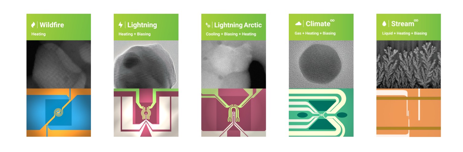
TEM relies on specialized MEMS chips and liquid or gas micro-cells, while SEM typically uses environmental chambers and nanoindenters
A lab studying atomic diffusion in a nanoparticle needs TEM's resolution. A lab studying how a coating fractures under mechanical load is usually better served by SEM. Neither is a lesser choice, they answer different questions, and it's usually the first thing Sigma's application team works through with a research group before recommending a specific In Situ setup.
How the hardware makes it possible
This is where the engineering gets specific, and where DENSsolutions has built a name across research labs handling In Situ TEM work. Rather than one general-purpose holder, DENSsolutions' range is built around the type of stimulus a study needs:
1. Heating studies up to 1300°C with stable sample positioning
2. Combined gas, heating and biasing work for catalysis and fuel cell research, with data correlation between structural and chemical results
3. Liquid-phase experiments run through a sealed nano-cell, useful for electrochemistry studies across a range of temperatures
4. Combined electrical biasing and heating through a four-point-probe chip design, for studies that need both stimuli applied at once
None of this replaces sound experimental design. A well-built holder still needs a researcher who knows which stimulus, which temperature range and which resolution actually answers their question, which is where Sigma's own application specialists tend to get involved early, well before a DENSsolutions holder is even installed. But the equipment itself does mean the microscope stops being a still camera and starts behaving like a live laboratory.
Where Sigma fits into this
Sigma Enterprises has worked across material science and NDT inspection instrumentation for close to two decades, supporting university and industrial research labs across the UAE and GCC. DENSsolutions sits within that portfolio, alongside microscopy names like Evident and WITec, as part of a broader materials characterization offering rather than a single-product line, and Sigma's team has spent years getting familiar with exactly where DENSsolutions equipment fits against the alternatives.
For a lab evaluating In Situ TEM equipment, that usually means the conversation with Sigma doesn't end at the sale. Application support, on-site installation, calibration and a regional service center matter as much as the specification sheet, particularly for equipment this specialized, where a delay in support can stall an entire research timeline. It's this ongoing relationship, not just the DENSsolutions hardware on its own, that Sigma positions itself around.
The bigger question worth sitting with isn't whether In Situ imaging is useful. The results across catalysis, battery and corrosion research already answer that. It's whether a lab's current setup is still asking researchers to infer what happened, when it could be showing them instead.